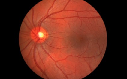
Retinal detachment is one of the few eye emergencies. There are three general types of retinal detachment. All three forms have an elevation or a lifting off of the retina itself off the inside back wall of the eye, with an accumulation of fluid between the retina and the eye wall. The retina is the lining or “wall paper” of the back of the eye. The retina covers the inside of the eye and contains all the sensors that detect light and send images to the brain for us to process. The most critical area to vision is the macula which is an area of the retina.
The symptoms of a retinal detachment are: flashes of light, an increase in floaters, a curtain or shadow moving over the field of vision in the center or peripheral vision, and finally total loss of vision peripherally and /or centrally. If you experience any of these symptoms you should call your ophthalmologist and /or go to the emergency room.
The first type of retinal detachment is rhegmatogenous, which is an elevation of the retina and a flap tear or break in the retina. The elevation is usually fluid that does not shift with body position. The second type, an exudative retinal detachment,causes minimal-to-severe visual loss or visual defects. This type also has serous fluid elevation of the retina and the fluid does shift when the person changes position. Chronic inflammatory diseases, congenital eye abnormalities, can cause this fluid accumulation and unknown causes classified as idiopathic. The third classification, tractional retinal detachment, may be symptom free or the person may have some vision loss or defect. This final type can be caused by bands of “cob-web-like” traction bands that form in the back of the eye and contract and pull the retina off the eye wall. The causes of these bands are diabetic retinopathy, sickle-cell retinopathy, retinopathy of prematurity, and trauma.
The treatment of a person with an “acute” rhegmatogenous retinal detachment that involves the macula or those with tractional retinal detachment that involves the macula is bed rest and surgical repair performed as soon as possible. All other retinal detachments that do NOT threaten the macula are repaired at the earliest convenience, preferably within 1-2 days. Chronic retinal detachments are usually treated within one week. For the exudative retinal detachment, the successful treatment of the underlying condition often leads to resolution of the detachment. This is a basic overview of retinal detachment and for further questions consult your ophthalmologist.